
The webinar. Now with every answer.
Watch the recording, read the complete answers from the Q&A and choose the next step that fits your practice.
Webinar recording
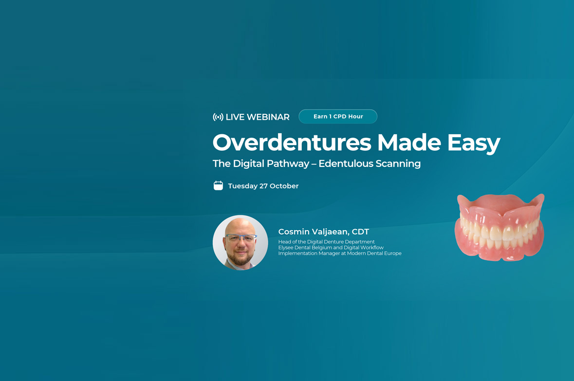
The webinar you registered for.
Chapter markers take you directly to accuracy, workflow, support and ROI.
Your questions from the webinar
The main participant questions, edited for clarity and available for reference.
-
What should we do if pronounced undercuts prevent us from accurately scanning a relined denture?
A reline records the current supporting tissues and may introduce undercuts that are difficult to capture with a clinical scan. Two workflows were described.
Option 1 Send the relined denture. Reline the existing denture and send it to the
laboratory for a complete 360-degree scan. The laboratory then returns the denture. The patient may be without it for approximately one day, depending on the local laboratory and transport distance.Option 2 Use a printed duplicate. Scan the existing denture through 360 degrees before relining and send the scan to the laboratory. The laboratory can print a duplicate try-in for you to reline. The relined duplicate can then be digitised in the laboratory, allowing the patient to retain the original denture throughout the process.
-
When changing the vertical dimension of occlusion, should we scan at the new position, and how can we stabilise the occlusion during scanning?
Ideally, record the intended vertical dimension of occlusion (VDO) directly in the patient and scan at that position. If the existing occlusion is submitted, the laboratory can increase the articulator pin height, for example by 2-3 mm, but this does not reproduce the patient’s individual hinge-axis relationship.
- Use wax between the arches or a premeasured rubber jig between the anterior teeth to maintain the intended opening. Jigs with thicknesses in 1 mm increments, up to approximately 4-5 mm, were described.
- With the position stabilised, take the two lateral bite scans to record the revised occlusal relationship.
- State clearly on the laboratory prescription that the VDO was adjusted clinically and that the scanned relationship should be used as supplied.
This instruction prevents uncertainty when the laboratory sees an increased opening or a lack of tooth contact and would otherwise need to request confirmation.
-
In the protocol presented during the webinar, why is the impression taken in the try-ins rather than in the occlusal rims?
The try-ins provide an established, stable occlusal relationship that can be used to control their position during a functional impression. This is more difficult when an occlusal rim is held in position by hand.
During an impression or reline of an upper denture, a lack of occlusal control can allow the denture to tilt posteriorly at the A-line. The speaker illustrated this with approximately 0.5 mm of material anteriorly near the incisive papilla and 2-3 mm posteriorly. Such an uneven thickness can reflect displacement of the denture during the impression.
Taking the impression with the opposing try-ins in occlusion helps control this tilt and allows the impression to be functionalised while maintaining the established relationship. This recommendation arose from discussions with clinicians about the difficulties encountered in practice.
-
How long are the digital files retained, and why are they useful if the supporting tissues change over time?
The stated target is to retain the digital files for 10 years.
The supporting tissues may change after one or two years, but the stored denture design remains useful. It preserves the original tooth arrangement, aesthetics and occlusal relationship. The fitting surface can subsequently be adapted through a reline while retaining those design features, even several years later; five years was given as an example.
The purpose of retaining these records is to support continuity for the patient and reproduce a familiar appearance. A denture is a prosthesis, and patients may value being able to maintain their established appearance rather than having visibly different teeth each time a replacement is required.
The speaker emphasised that successful dental technical work can be unobtrusive:
preserving a natural, familiar result is an important reason for keeping the original digital designs. -
What workflow is recommended when making a new overdenture on an existing bar that we want to retain?
The recommended workflow combines a digital design with a conventional functional impression. Although the prosthetic area and existing bar can be scanned to design the overdenture, the impression is used to refine the fit to the supporting mucosa.
- Scan the prosthetic area and existing bar so that the laboratory can design the
overdenture and provide a try-in. - The laboratory provides sufficient space in the try-in for impression material. Use the try-in to take a functional impression in occlusion and return it to the laboratory.
- The laboratory produces a model from the impression and incorporates the new model into the digital design. If the occlusion and aesthetics have been approved, these are retained while the fitting surface is adapted to the updated model.
- The laboratory can then incorporate the attachments and complete the denture in the final production stage.
The rationale is to retain the benefits of digital design while using a conventional impression where it provides greater precision for the fit to the supporting tissues.
- Scan the prosthetic area and existing bar so that the laboratory can design the
-
Can Evo Fusion dentures and overdentures be made with metal reinforcement?
Yes. The options discussed included mesh reinforcement and internal metal frameworks.
For example, in a maxillary overdenture retained by four Locator attachments, an internal framework can be covered by the denture base. This avoids exposed metal on the surface contacted by the tongue for patients who dislike that sensation.
Digital design allows the laboratory to control the framework’s shape and thickness. The speaker noted that waffle-type reinforcement is used less frequently for lower dentures when an internal framework can be designed, but it can still be incorporated if requested by the dentist.
The reinforcement design should be discussed with the local laboratory according to the case requirements and the dentist’s prescription.
-
Further questions and laboratory support
For questions about a specific case or assistance with the workflow, please contact your local Modern Dental laboratory or branch. The laboratory team can advise on practical difficulties encountered during digital denture and overdenture treatment.
What we learned from the webinar
Complete Digital Workflow
Plan a complete digital workflow for edentulous patients.
Predictable Scanning
Apply a predictable scanning strategy for soft tissue and feature-poor anatomy.
Accurate Attachment Capture
Capture bars and locator attachments accurately for digital design.
Bite Registration & Jaw Relations
Record reliable bite registrations and jaw relations while avoiding common errors.
Digital Design & Production
Understand the digital design and production workflow for overdentures and implant-retained prostheses.
Treatment Efficiency
Improve treatment efficiency, reproducibility, and patient outcomes through digital workflows.
Choose your next step
Price List & Brochure
If you would like to receive our price list and product brochure click on the relevant button below.
Request a Call back
Fill out your contact details and a member of our team will get in touch.
Sign-up to our Newsletter
If you would like to receive our monthly updates via our newsletter click on the button below.